In a Dog's Eye
The visual system is an important sense available to the canine. The eyeball functions to gather light and transmit it into electrical impulses which are interpreted by the brain to form images.
The visual system is an important sense available to the canine. The eyeball functions to gather light and transmit it into electrical impulses which are interpreted by the brain to form images. The eye is protected by being sealed in a bony eye socket supported by lubricating tissues, muscles, and eyelids.
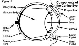
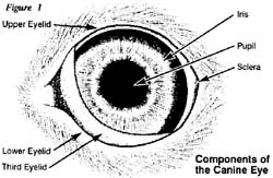 The eyeball itself is formed by layers of tissue (see Figures 1 & 2). The white area called the sclera is made of tough fibers rich in blood vessels which transport oxygen and nutrients to the eye. The clear portion of the eye is the cornea and it is made of layers of cells arranged in a unique fashion so as to be transparent. The cornea lets the light enter in to the eye. Inside the eyeball are specialized organs bathed in a liquid called vitreous fluid, which keeps the eyeball "inflated" and supplies nutrients to the structure within the eye. In the interior of the ball is the colored portion called the iris. As in humans, dogs have different colored eyes and this is determined by the color of the iris. In the center of the iris is an opening called the pupil. This opening can be made larger or smaller as muscles called ciliary bodies ( which are attached to the colored iris ) expand or contract. In dim light, the pupil is made larger so as to let more light enter the eye. Conversely, in bright light the pupil becomes smaller. This is important as too much can cause pain and/or damage to the eye just as in humans.
 Behind the pupil lies the lens which is a pea sized organ that is normally clear. The lens bends, concentrates and focuses the light so it will land on the rear area of the eyeball called the retina. The retina contains nerve cells called rods and cones. The rods are sensitive to light and the cones are sensitive to color. Unlike humans, the canine possesses very few cones and that is why domestic dogs are thought to be color blind. They see only shadow of gray, black, and white. Dogs do, however, have many rods and other reflective cells which enable them to see in very dim light. The nerve cells within the retina transform the light into nerve impulses which leave the eyeball by the optic nerve and enter the brain. The brain translates the impulses into images, creating vision.
Several eye disorders have been identified in the Jack Russell Terrier. This article touches on only some of the ones that have come to attention through recurrence. They vary in severity from those that progress with a few, subtle signs , like PRA ( Progressive Retinal Atrophy ), to those that strike like a thunderclap, like PLL ( Primary Lens Luxation ), when the dog is fine Thursday and blind on Friday. With the help of the general membership in testing of their own dogs, more information can be gathered in the quest to identify and eliminate these problems.
Cataracts
A cataract is defined as a loss of the normal transparency of the lens of the eye. Any spot on the lens that is opaque, regardless of size, is considered a cataract. They may affect one or both eyes. Some are clearly visible to the naked eye, appearing as white or bluish dots.
Treatment consists of surgical removal of the lens ( cataract extraction ). This operation is usually recommended for the dog who has so much visual impairment that it has trouble getting around.
The prudent approach is to assume cataracts to be hereditary. However, cataracts may result from injuries to the eye, exposure to great heat, radiation, diabetes, and old age (senile). Cataracts can also occur from ocular inflammation, specific metabolic diseases, persistent pupillary membrane ( PPM ), or nutritional deficiencies.
Breeding of affected animals is not recommended.
Distichiasis
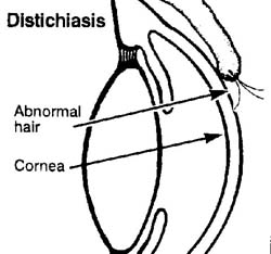 Distichiasis is a condition in which small hair structures abnormally grow on the inner surface of the eyelids ( see diagram ). Both upper and lower lids may be involved. The abnormal hairs growing on the inner surface of the lids cause irritation to the cornea. The affected eye will become red, inflamed, and may develop a discharge. The dog will squint or blink very often, much like a person when a bug or other foreign matter enters the eye. In severe cases, the cornea may become ulcerated and appear bluish in color. Left untreated, severe corneal ulcerations and infections can develop. The hairs can cause severe irritation and without treatment will usually worsen. Blindness can result if infections do develop.
Treatment involves the removal of the hairs through the use of surgery or electro-epilation. With electro-epilation, a fine needle is passed into the hair follicle and the follicle is burned to destroy the hair and its roots. If surgery is performed, the lid is actually split and the areas with abnormal hairs are removed. Antibiotic eye drops will also be used with the surgery to eliminate infections.
Glaucoma
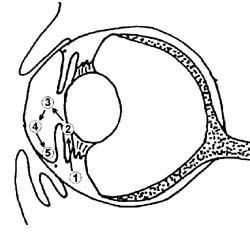 This is a serious eye problem. Usually it leads to partial or total blindness. It is due to an increase of fluid pressure within the eyeball caused by an interruption of fluid exchange between the eyeball and the venous circulation. The fluid that develops the excessive pressure associated with glaucoma is the aqueous humor, the liquid found in front of the lens. It is produced by the ciliary body (1), flows past the lens (2), through the pupil (3), across the inner surface of the cornea and front side of the iris (4), and then drains from the eye at the angle ( commonly referred to as the drainage or iridocorneal angle ) formed where the cornea meets the iris (5). The cornea and the lens are living tissues but they have no blood vessels going to them to supply the needed oxygen and nutrition. These life sustaining materials are brought to them through the aqueous humor. In glaucoma, elevations in the pressure of the aqueous humor are most frequently caused by this fluid not being able to drain correctly from the eye. When the eye pressure is greater than the arterial blood pressure, arterial blood cannot enter the eye to nourish the retina. A sudden build up of pressure leads to acute blindness and damage to the optic nerve. A slower build up causes few symptoms, yet leads to the same results.
Primary (or congenital) glaucoma occurs without prior disease. It occurs in a dog because it possesses physical or physiologic traits that predispose it to glaucoma. These are usually predetermined by genetics. Secondary glaucoma means that the disease is secondary to, or caused by, another condition. A common example is a penetrating wound to the eye. Other causes would include bleeding in the eye, inflammation within the eye, attachments or scarring between the iris and lens, and luxation or displacement of the lens. Lens luxation can cause glaucoma and likewise chronic glaucoma can cause lens luxation. The early sings of glaucoma that an owner may watch for or notice are pain, a dilated pupil, cloudiness within the cornea and /or an increase in the size of the blood vessels in the white portion of the eye. The pain may present itself with the dog rubbing his eye with his paw, against the furniture or carpet, or your leg. This is a common, and often unnoticed, early sign. Some dogs will also seem to flutter the lids or squint with one eye. The pupil of the affected eye will usually dilate early in the course of the condition. It may still react to a bright light shining in it, but it will do so very slowly. Remember that glaucoma, even primary glaucoma, is usually going to initially affect just one of the eyes. If the pupil in one eye is larger than the other, something is definitely wrong.
Just so there is no misunderstanding, if treatment in the dog is not started to combat glaucoma in a few days or, in some cases a few hours, vision will probably be lost completely from the affected eye. The pressure can crush the cells of the retina and optic nerve, rendering them nonfunctional. It can break down the structures holding the lens in place and it can cause damage to the iris and cornea. After these internal changes have occurred, the eyeball itself swells in size, tilts off to the side, and all the surface blood vessels enlarge giving it the appearance of a large, ugly, bruised radish.
Measurement of the intraocular ( inner eye ) pressure and interior inspection of the eye is needed for diagnosis. Chronic glaucoma can be managed for a time with drops and medication.
Breeding of affected animals is not recommended.
Luxated Lens
 Luxated (displaced) lens occurs when the zonula (ligament fiber) which holds the lens in place deteriorate allowing the lens to fall out of its normal site behind the pupil. Subluxation is the partial separation of the lens position and is often times an indication of eventual total luxation. When the lens luxates to the posterior (rear) chamber of the eye, the eye will appear normal, but if it luxates forward (anterior), the lens will rub against and irritate the cornea, causing tearing and a bluish cast over the eye. Anterior lens displacement is the most hazardous form of the displacement. It has a high probability of causing glaucoma, and if the lens touches the cornea it will cause damage leading to cornea edema. This must be attended to immediately as the lens now restricts the flow of ocular fluids, creating eye pressure and great discomfort. Before luxation is apparent, you may notice behavior changes in the dog; not catching a biscuit tossed to him, bumping into stationary objects, missing the first step in a staircase.
Treatment varies due to the severity of the disorder. Surgical removal of the lens will alleviate pain and allow partial vision. This surgery is expensive and not always shown to be effective. Sometimes a combination of eye drops and oral medication is helpful. In severe cases, removal of the eye is necessary.
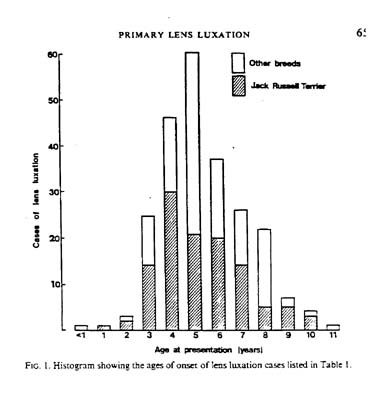
Primary lens luxation is assumed to be autosomal recessive. That is, each parent must at least be a carrier of the disorder. If a dog is affected, certain facts are known; both parents are at least carriers and every offspring of the affected dog is a carrier. Affected dogs should not be bred and known carriers should be pulled from the breeding program. This is an insidious disorder as it appears anywhere between 3-8 years of age, well into the breeding program. With primary lens luxation, at first only one eye is affected but it is only a matter of time before the other eye follows suit. The time can vary from several weeks to several years, but it will follow. Secondary lens luxation is associated to trauma to the eye, such as puncture or injury, and is not hereditary.
PPM
Persistent pupillary membranes (PPM) are blood vessel remnants in the anterior (forward) chamber of the eye which fail to regress normally in the neonatal period. These strands may bridge from iris to iris, iris to cornea, iris to lens, or form a sheet of tissue in the anterior chamber. The last 3 forms pose the greatest threat to vision and when severe, vision impairment or blindness may occur. The membranes appear to be white, gray, or pigmented, and do not usually involve >25% of the corneal surface. There is no effective treatment for PPM.
PRA-PRD
Progressive retinal degeneration ( PRD ) is also known as progressive retinal atrophy ( PRA ) and refers to retinal diseases that cause blindness. Some breeds have blindness by abnormal development of the retina and this is dysplasia. Other breeds have a slowly progressive degeneration or death of the retinal tissue and this is degeneration.
As the name PRD implies, a slow death of the retinal tissue occurs. It is a slowly progressive disease and the earliest signs may be overlooked. The first indication of a problem is the loss of night vision. The dog hesitates to go out at night. He won't jump on or off furniture in a darkened room. Later he will go up, but not down, stairs. A loss of day vision will follow.
The veterinary ophthalmologist examines the retina with an instrument called an indirect ophthalmoscope. Changes in the retinal blood vessel pattern, the optic nerve head, and the reflective substance within the dog's eye called the tapetum can be seen which are classic for PRD. However, in some breeds PRD characteristics have little or no early changes. The eyes of these dogs may appear normal until they are in the later stages of the disease. Progressive retinal degeneration will progress at different rates in different breeds. This variation causes difficulty in determining just how long any particular dog will continue seeing. Cataracts may occur in some patients with PRD and generally occur later in the disease. Formation of cataracts may interfere with the ophthalmologist's direct examination of the retina and make other tests such as an electroretinogram (ERG) essential for diagnosis. The ERG is sensitive enough to diagnose dogs with PRD before they begin to demonstrate signs of the disease.
In summery, PRD refers to a broad group of inherited retinal disease which result in the blindness of dogs. Because of the nature of the disease and sometimes late onset, repeated examinations may be required to detect individuals with the condition. Patients affected should not be used for breeding.
PRA has been shown to be autosomal recessive in the poodle, Irish setter, Norwegian elkhound, and Samoyed. Inheritance should be assumed in other breeds. The recessive nature makes this disease extremely difficult to eliminate from affected bloodlines. There is no treatment for the disorder.
In closing, let me leave you with some facts and figures from CERF, the Canine Eye Registration Foundation; from January 1991 to May 1996, a grand total of 364 Jack Russell Terriers were given eye examinations by board certified canine ophthalmologists. Of those, 80% were eye normal and 20% had some form of eye disorder. Of those with eye disorders, 50% were so severe as to be rejected for registration by CERF.
Now look at the total membership of the JRTCA and the number of registered dogs over that same period of time: probably close to 5000 dogs. Yet only 364 were examined by CERF. If we, as a club, are ever going to get a grip on the disorders, of all kinds, that affect our breed, we must start testing our dogs for everything we can: eyes, hearing, orthopedics, etc. This can't be stressed enough. It's up to us, the breeders, to make our breed as healthy and sound as we possibly can. Only we can do it. And if you profess your love of the breed, your desire to see it flourish, you must let your actions speak for you. Do the best you can, test your dogs, and pull those that fail. Breed only the best. If we all work together, we can make the Jack Russell Terrier a strong, healthy, defect free breed.
by Alice Reynolds
References
Bailliere's Comprehensive Veterinary Dictionary
D.C. Blood & Virginia P. Studdert
Canine Eye Registration Foundation
Breakdown report Jan. 1991- May 1996
Drs. Foster & Smith Canine News
Vol. 3 #6 June 1994
Vol. 3 #11 Nov. 1994
Dog Owner's Home Veterinary Handbook
Delbert G. Carlson D.V.M. & James M. Giffin, M.D.
Handbook of Small Animal Practice
2nd edition Rhea V. Morgan
Ocular Disorders Proven or Suspected to be Hereditary in Dogs
American College of Veterinary Ophthalmologists
Progressive Retinal Atrophy
Dr. Dennis Hacker
Veterinary Ophthalmology Notes
2nd edition Glenn A. Severin D.V.M., M.S.
by Alice Reynolds